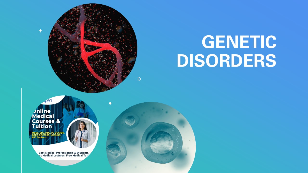
GENETIC DISORDERS MBBS NOTE
- The group of disorders affecting the foetus during intrauterine life (genetic as well as developmental) and paediatric age group.
- Groups of genetic diseases include :
- Developmental defects
- Cytogenetic (Karyotypic) defects
- Single-gene defects (Mendelian disorders)
- Multifactorial inheritance disorders
- Storage diseases (Inborn errors of metabolism)
Congenital Disorders
- Non Genetic:
- Developmental defects – Malformations
- Genetic Disorders
- Chromosomal
- Gene - Mendelian
- Multifactorial
- Human germ cells (ova and sperms) contain 23 chromosomesm(haploid or 1N)
- while all the nucleated somatic cells of the human body contain 23 pairs of chromosomes (diploid or 2N)— 44 autosomes and 2 sex chromosomes, being XX in females (46, XX) and XY in males (46, XY).
- Human germ cells (ova and sperms) contain 23 chromosomesm(haploid or 1N)
- while all the nucleated somatic cells of the human body contain 23 pairs of chromosomes (diploid or 2N)— 44 autosomes and 2 sex chromosomes, being XX in females (46, XX) and XY in males (46, XY).
- Chromosomal banding techniques are employed for study of classes of chromosomes.
- Chromosomal bands are unique alternate dark and light staining patterns.
- Banding techniques include:
- i) G-banding (Giemsa stain);
- ii) Q-banding (quinacrine fluorescence stain);
iii) R-banding (reverse Giemsa staining); and
- iv) C-banding (constitutive heterochromatin demonstration).
- Cytogenetic disorders- structural (nondisjunction during gametogenesis, numerical-(from error during division)
- Monosomy – associated with one less normal chromosome( 2n-1)
- Trisomy- one extra chromosome( 2n+1)
- Mosaicism( Mitotic errors in zygote)- associated with 1 or more population of cells, some normal chr., others with extra or missing chr.
- Mendelian (single gene mutation)
Mutations
- Genome: whole set – Polyploidy 4n, 8n etc.
- Chromosomal: change in chromosome.
- Number: Trisomy, monosomy
- Structure: Deletion, Translocation etc.
- Gene: Submicroscopic.
- Point mutation – single base sequence
- Deletions -
- Insertions
Cytogenetic Abnormalities:
- Abnormal number of chromosomes:
- Non-disjunction - Down’s Syndrome
- Anaphase lag - Turner’s xxx
- Abnormal Structure: (normal no)
- Deletion of short arm 5q- Cri-du-chat syndrome
- Inversion -
- Translocation - Ph Chromosome - t(9:22) CML
TURNER SYNDROME
- Hypogonadism in Phenotypic females
- Complete or partial Monosomy of X Chr.
- 45,X most common
- Mosaics also occur (eg., 45,X/46,XX)
Structural abnormalities of X chromosomes
- Deletion of the small arm, resulting in the formation of an isochromosome of the long arm
- Deletion of the proportions of both long & short arms resulting in the formation of a ring chromosome
- Deletion of portions of the short or long arm
KLINEFELTER SYNDROME
- Male hypogonadism
- 2 or more X chromosomes and at least one(1) Y chromosome.
- 47,XXY most common(80%)
- Mosaics also-(e.g., 46,XY/47,XXY)
CLINICAL FEATURES
- Commonest cause of male infertility
- Body Habitus- Eunuchoid
- Failure of Male secondary sexual character
- Gynecomastia
- Female distribution of hair
- Atrophic testis with hyperplasia of leydig cells
- FSH & Estrogen levels elevated, testosterone levels low
- Minimal or no Mental retardation
DOWNS SYNDROME (Trisomy 21)
- Most common 1 in 800 births
- Complete extra chromosome 21(47,XY,+21)
- Increased maternal age -1 in 25 above 45 years of age
- Cause is meiotic non disjunction of chromosome 21 in the ovum
- Mosaic variant 1%
Clinical features
- At birth evident
- Flat facial profile
- Oblique palpebral fissures
- Epicanthic folds- “Mongolism”
- Mental retardation- 80%
- Shy & Gentle!
- Mosaics- Near normal intelligence
- 40% congenital heart disease- ASD, VSD, AV valve malformations
- Atresias of esophagus, small bowel
- 10 to 20 fold increased risk of- Acute leukemias (AML, ALL)
- All above 40y develop Alzheimer disease
- Abnormal immune response- Predisposed to serious infections in lungs, thyroid autoimmunity
SINGLE-GENE DEFECTS (MENDELIAN DISORDERS)
- The classic laws of inheritance of characteristics or traits were outlined by Gregor Mendel in 1866.
- Single-gene defects follow the classic mendelian patterns of inheritance and are also called mendelian disorders.
- These disorders are the result of mutation of a single gene of large effect.
ENZYME PROTEIN DISORDERS OR LYSOSOMAL DISORDERS
Mutation of gene
Alters the protein synthesis
Metabolic block
NORMAL METABOLISM
Enzyme act as catalyst
In case of deficiency
accumulation of intermediates
decrease in end product
no inactivation of tissue damaging sub
PHYSIOLOGY OF LYSOSOMES (HYDROLYTIC ENZYMES)
- Synthesized in ER
- Compelled in Golgi
- complex with M 6 P
- binds to receptors
- separated as vesicles
- Fused with lysosomes
- Digests the macromacules
- metabolic intermediates
- intracellular organelle
- phagocytosed material
PATHOLOGY OF STORAGE DISORDERS
- Abnormal enzymes
- No proper digestion (phagocytosis, organs, metabolism)
- Increase accumulation in the lysosomes
- Interfere in normal cell function
- C/F depends on site of enzymes
- glycosidase ---- neuron
- muco polysaccharide-----every organ
- Rich in Mononuclear Phagocytic cells (MPC) which degrades many substance
- MP Cells are rich in liver, spleen, LN, Marrow etc.
CLASSIFICATION OF L.S. D
- GLYCOGEN STORAGE DISORDERS
- Von gierkes
- Pompe
- SPHINGOLIPIDOSES(GANGLIOSIDOSIS)
- GM1 gangliosidosis( inf, juv)
- GM2 (Tay sachs, Sandhoffs)
- SULPHATIDOSES
- Gouchers
- Niemen picks
- MUCOPOLYSACHARIDOSIS
- 6 types
- MUCOLIPIDOSES
- OTHER
VON GIERKES DISEASE
- Glycogen start accumulating
- No glucose release
- Hypoglycemia
- Increase fat metabolism
- Deficiency of Glucose 6 Phosphatase
- Increase lactate, pyruvate & ketosis
- C/F Childhood
- hepatomegaly
- failure to thrive
- skin xanthoma
- cytoplasm clear glycogen
POMPES DISEASE
- Decrease in acid maltase
- Glycogen accumulation in heart and muscle
- C/F cardiomegaly, cardiorespiratory failure
- Hypotonia and muscle weakness
- Die within 2 years
- In adult form mainly skeletal muscle involved
TAY SACHS DISEASE
- Common type o f GM2.
- Mutation of Ch 15. hexosaminidase deficiency
- Gangliosides accumulates in
- liver, spleen, heart , CNS, Retina.
- Ballooning of neurons
- cytoplasm filled with gangliosides
- cytoplasmic vacuoles
- special stain oil red O, Sudan black
- EM vacuoles showing onion skin appearance
TAY SACHS DISEASE
- Progressive destruction of neurons
- Proliferation of microglia.
- Involve cerebellum, basal ganglia, ANS, cord.
- Involve the MPC.
- Retina shows cherry red spot
C/F OF TAY SACH
- Varies depending on the degree of deficiency
- Normal at birth
- By 6 month motor and mental deterioration
- muscle flaccid
- motor in coordination
- Mental
- dementia,
- blindness
- pathetic
- cherry red spot.
TAY SACHS DISEASE
Diagnosis
- enzyme assay
- DNA analysis
NIEMANN -PICK DISEASE
- Accumulation of sphingomyelin & cholesterol
- Deficiency of sphingomyelinase
- Sphingomyelin present in cell and organeller membrane
- No degradation of membrane
NIEMANN -PICK DISEASE
TYPE A
- severe and infantile form
- extensive neuronal involvement
- viscera also accumulated with SM
- progressive disease
- death with in 3 yrs.
NIEMANN -PICK DISEASE
TYPE B
- CNS not involved
- adult form
- organomegaly
MORPHOLOGICAL CHANGES
- TYPE A
- Absence of sphingomyelinase
- accumulation of sphingomyelin in MPC
- Cells are enlarged, distended lysosomes
- filled with sphingomyelin and cholesterol
- small uniform, multiple vacuoles & foamy cytoplasm
- frozen section & special stain for fat.
- EM Shows zebra bodies
MORPHOLOGICAL CHANGES
TYPE A
- lipid laden cells in spleen, LN, marrow, GIT, lung.
- splenomegaly up to 10 times,
- moderate hepatomegaly, LN enlargement.
- Brain sulci widen, shrunken gyri.
- all part of nervous system are involved.
- Neurons are ballooned, and decreased in number.
- Cherry red spot in the retina.
CLINICAL FEATURES OF NIEMANN- PICK
- Start at birth
- Evident by 6 months
- Hepato splenomegaly and failure to thrive
- Lymphadenopathy
- Fever
- Vomiting
- Death within 2 years
DIAGNOSIS OF NIEMANN- PICK
- Sphingomyelinase assay in liver & marrow.
- DNA probe analysis
GAUCHERS DISEASE
- Glucocerebrosidase deficiency
- Glucocerebroside accumulation in lysosomes
- Present in phagocytic cells and CNS
- It is the most common disease
- Cluster of Autosomal recessive disease
- There are 3 types
TYPE 1 GAUCHERS (CLASSIC/ADULT FORM)
- 80% of Gauchers
- Glucocerebrosides accumulates in the MPC
- Spleen, liver, bone marrow, lymph nodes and skeletal muscles are involved
- Brain is not involved.
- Life is short but not marked
TYPE 2 GAUCHERS (INFANTILE FORM)
- Neuropathic form
- Progressive involvement
- No detectable glucocerebrosidase
- Hepato splenomegaly
- Patient die in early age
TYPE 3 GAUCHERS (JUVENILE FORM)
- It is between 1 and 2
- Progressive involvement of CNS
MORPHOLOGY OF GAUCHERS
- Massive accumulation in the MPC all over
- Gauchers cells liver, spleen, marrow, LN
- Fibrillar type of cytoplasm
- Crumpled tissue paper appearance.
- Cells up to 100u
- Eccentric nucleus
- PAS positive
- EM Elongated lysosomes with lipids
- Spleen enlarged to 10 times
- Pale and matted surface (focal accumulation)
- Moderate lymphadenopathy
MORPHOLOGY OF GAUCHERS
- BONE
- Erosion
- skeletal deformity
- soft and gray
- pathological fracture
- CEREBRUM
- Gauchers cells in the virchow robins space
- No lipids in the neurons
- Neurons are destroyed possible toxic effects of surrounding cells
CLINICAL FEATURES Type 1 Gauchers
- Adult type
- Pancytopenia due to hypersplenism
- Pathological fracture
- Bone pain
- Compatible with long life
TYPE 2 AND 3 OF GAUCHERS
- Convulsion
- Mental deterioration
- Liver, spleen and LN are affected
MUCOPOLYSACHARIDOSIS
- Is a group of disorders
- Enzymes degrading mucopolysacharides
- MP are glycosamino glycans or lecithin complex COH linked to proteins.
- Accumulation of dermatan, keratan, heparan and chondroitin sulphate.
- The enzymes cleaves terminal sugar and allow to digest further.
MUCOPOLYSACHARIDOSIS cont….
- Abnormalities in enzymes in the lysosomes
- Accumulation of substance in the lysosomes
- Cause somatic and neurological problems
- There are 7 types
- Hunter is X linked and all other are Autosomal
- Progressive disorders of multiple organs involves liver, spleen, heart and vessels.
MORPHOLOGY OF MUCOPOLYSACHARIDOSIS
- Substance accumulates in the MPC, endothelium and SMC.
- It involves the spleen, liver . LN, heart and vessels.
MICROSCOPY OF MUCOPOLYSACHARIDOSIS
- The cells are ballooned with mucopolysacharides.
- Cytoplasm have many clear vacuoles
- EM shows swollen lysosomes filled with granular PAS positive materials.
- Presence of zebra bodies in lysosomes
CLINICAL FEATURES
- Hepato splenomegaly and skeletal deformities.
- Sub endothelial deposition cause myocardial ischemia and MI.
- HURLERS
- Alpha 1 iduronidase deficiency
- Hepato splenomegaly
- growth retardation
- death in 6 to 10 years due to CVS complication
- HUNTERS
- It is very mild disease
- corneal clouding
TUMORS OF INFANCY AND CHILDHOOD
- Tumors and Tumor-like Conditions
- MC neoplasms of childhood = soft-tissue tumors (mesenchymal) - adults
- Benign tumors more common than cancers
- 2% of all malignant tumors occur in infancy and childhood
- Tumor-like conditions
- Heterotopia (or Choristoma )
– Normal cells or tissues that are present in abnormal locations
– pancreatic tissue in stomach or small intestinal wall
– adrenal cells found in the kidney
- Hamartoma
– Excessive, focal overgrowth of cells / tissues native to the organ but not having normal architecture
– Examples = Adenomas of the liver, Hemangiomas, lymphangiomas, rhabdomyomas
Benign Tumors
- Hemangioma = MC tumors of infancy
– Cavernous and capillary types
– Common on face and scalp
– Flat , larger lesions = port-wine stains
– Hereditary disorder:- von Hippel – Lindau (VHL) disease.
- Lymphatic tumors
- Lymphangiomas (hamartomatous or neoplastic)
– increase in size after birth
- Encroach on vital structures in mediastinum or nerve trunks of axilla
- Lymphangiectasis = not progressive,
- Fibrous tumors
- More common =Fibromatosis, sparsely cellular (multiple fibromas)
- Congenital - infantile Fibrosarcomas {t (12:15)}
– ETV6-NTRK3 fusion transcript = diagnostic marker
– ETV6 (transcription factor) and NTRK3 gene is tyrosine kinase.
- Teratomas
- Histological maturity correlates with biologic behavior
- well-differentiated cystic lesions (mature teratomas),
- Age = two peaks ( 2 years of age & late adolescence or early adulthood)
- Sacrococcygeal teratomas = MC ( more common in girls (M:F::4:1)
- 75% are mature and benign teratomas
- Other sites of teratomas =testis, ovaries, midline
Malignant Tumors
– Incidence and types
– Neuroblastic tumors
– Wilm’s tumor
- Neuroblastic tumors-
- Tumors of the sympathetic ganglia and adrenal medulla
- Derived from neural crest cells
- Neuroblastoma
– MC extra cranial solid tumor of childhood
– median age at diagnosis = 18 months
– MC diagnosed tumor of infancy
– Most of them are sporadic
- Most characteristic features
– spontaneous or therapy-induced differentiation of primitive neuroblasts into mature elements
– spontaneous tumor regression
– wide range of clinical behavior and prognosis,
- Children younger than 18 months of age have better prognosis
- 5-year survival is generally 40%
Malignant = Neuroblastic tumors contd..
- Morphology
- Site = 40%arise in the adrenal medulla (MC site) followed by paravertebral region of abdomen (25%), posterior mediastinum (15%) and other sites (pelvis, Neck, brain (cerebral neuroblastomas)
- Size = minute nodules (in situ lesions) to 1 kg in weight
– Larger tumors have necrosis, cystic softening, and hemorrhage
– In situ ones are more frequent and spontaneously regress
- Histologically
- LM= Made of small, primitive cells with dark nuclei, scant cytoplasm, and poorly defined cell borders ; Karyorrhexis and pleomorphism are prominent; rosettes (Homer-Wright pseudorosettes)
- IHC =positive immuno –markers ( neuron-specific enolase (NSE), Synaptophysin, Chromogranin A, S100 protein etc.,)
- EM =cytoplasmic catecholamine-containing secretory granules with peripheral halo (dense core granules)
– Some show signs of maturation into ganglioneuroblastoma and ganglioneuroma
– Maturation is spontaneous or therapy-induced
– Differentiated lesions are accompanied by Schwann cells.
- Metastases to lymph node or/and liver, lungs, bone marrow, and bones
Malignant = Neuroblastic tumors contd..
- Staging.
- Stage 1: Localized with complete gross excision
- Stage 2: Localized with incomplete gross resection
- Stage 3: Unresectable unilateral, across the midline
- Stage 4: distant metastasis
- Stage 4S ("S" = special): infants younger than 1 year & dissemination limited to skin, liver, and/or bone marrow
- Clinical Course and Prognostic Features
- Under age 2 years= present with large abdominal masses, fever, weight loss
- Older children = insignificant until metastases; present proptosis (common metastatic site) and ecchymoses
– multiple cutaneous metastases ="blueberry muffin baby"
– 90% of neuroblastomas =produce catecholamines, helps in diagnosis (hypertension is less frequent ) vanillylmandelic acid [VMA] and homovanillic acid [HVA]) in urine or blood
- Prognosis
- "core" criteria used for risk stratification and therapeutic decision =age, stage, NMYC status, histology, and DNA ploidy
Wilm’s tumor
- MC primary renal tumor of childhood
- Peak age =between 2 and 5 years
- Improvements in the cure rates
- Pathogenesis and Genetics
- Increased association with four syndromes
– 1. WAGR syndrome =aniridia, genital anomalies, mental retardation; deletions of 11p13 (WT1 gene)
– 2. Denys-Drash syndrome= higher risk for Wilm's tumor (∼90%); gonadal
dysgenesis (male pseudohermaphroditism) and early-onset nephropathy; dominant-negative missense mutation in the zinc-finger region of the WT1 gene and increased risk for gonadoblastomas (WT1=critical for normal renal and gonadal development)
– 3. Beckwith-Wiedemann syndrome (BWS) ="WT2”gene
Genomic imprinting ; overexpression of IGF-2 (embryonal growth factor) is critical ; Organomegaly, macroglossia, hemi-hypertrophy, omphalocele, adrenal cytomegaly;
– 4. β-catenin associated Wilms tumors ; belong to WNT (wingless) signaling pathway; 10% of sporadic cases, gain-of-function mutations
- Nephrogenic rests =precursor lesions of Wilms tumors
- Clinical Features =large abdominal mass with hematuria, abdominal pain, intestinal obstruction and HTN
- Pulmonary metastases
- Prognosis depends on (poor prognostic features)
– Anaplasia,
– loss of genetic material on chromosomes 11q and 16q,
– gain of chromosome 1q
- Risk of Second malignancies
– soft-tissue sarcomas,
– leukemia and lymphomas,
– breast cancers
1 stromal -Usually fibrotic or myxoid in nature with Paler type of cells
2 blastemal - Bunch of less undifferentiated
- Dark cells Less differentiated therefore worse prognosis
3Epithelial -Form of abortive tubules or glomeruli as epithelial rosettes
- Better differentiated, therefore better prognosis.
................................................................................................
Coaching for 'Mbbs Students', 'Bds Students', 'Mds Students', 'Bams Students', 'Bpt Students', 'Bhms Students', 'Nursing Students', 'PreMed Students', 'Md Students', 'Doctor Students'
Connect with Bluepenonline
"Mbbs Tuition" "Bds Tuition" "Bams Tuition" "Next Tuition" "NeetPg Tuition" "Bpt Tuition" "PreMed Tuition" "Md Tuition"v"Medical Subjects Tuition" & "Medical Lecture Notes"
Please search in google or edge as follows to connect with Bluepenonline, Find Private & Affordable MBBS Tutoring, MBBS TUTORS, Online MBBS Tuitions, online classes for mbbs students, MBBS Tuition near me, BDS Tuition, Online BDS Tuitions, BDS Tuition near me, pre-med tuitions for international students, Online BAMS tuition, BAMS Coaching classes near me, BAMS coaching classes online, Online BAMS Coaching Classes, Next/NeetPg Coaching, ayurveda tuition, Ayurvedic medicine courses,..etc

 Common challenges faced by Ayurveda Students
 Common challenges faced by BPT & Nursing Students
 Common challenges faced by Dental Students
 Common challenges faced by MBBS Students
 What does an MBBS student learn?